Nano-Biotechnology
Keywords: Artificial Bioconstructs, High-resolution Imaging, Molecular Genetics, Nanodiagnostics, Nanostructured Surfaces, Organ-on-chip Technologies, Smart Biomaterials, Stimuli-responsive Carriers, Precision Medicine
The research lines of the Nano-Biotechnology area are listed below:
- Active Matter
- Biomolecular Delivery
- Biosensors & Biointerfaces
- Cellular and Molecular Biology
- Lab on Chip (LoC) & Organ on Chip (OoC)
- 3D Cell Models & Regenerative Medicine
- Advanced Imaging
Keywords: Active Matter, Soft Matter, Synthetic Biology, Optical Trapping, Holographic Microscopy
People: Silvio Bianchi, Roberto Di Leonardo, Claudio Maggi, Filippo Saglimbeni
Active matter encompasses all those systems composed by self-propelling constituents. In particular this class of systems include biological and synthetic micro and nano-swimmers. All these microparticles are capable of taking up energy from their environment and converting it into directed motion. Because of this constant flow of energy, their behavior can be explained and understood only within the framework of nonequilibrium physics. In the biological realm, many cells propel themselves to search for nutrients. These cells can genetically engineered to modify their collective behavior.
For instance the swimming speed of E. Coli can be modulated thanks to light-powered transmembrane proteins that forms the proton gradient exploited by the cell to spin their flagellar motors. By using light-shaping techniques it is possible to efficiently achieve spatial and temporal control of the cells’ speed in a bacterial bath. The spatial control of speed can be exploited to modulate the cell concentration and form a desired pattern (see figure above).
Keywords: Nanomedicine, Nanocarriers, Nano-Delivery, Nanoparticles, Stimuli-Responsive, Cancer Therapy, Gene Therapy, Plasma Medicine
People: Francesco Calabi, Eliana D’Amone, Stefania D’Amone, Pietro Favia, Roberto Gristina, Clara Guido, Stefano Leporatti, Georg Mahlknecht, Ilaria E. Palamà, Fabio Palumbo, Elisabetta Perrone, Francesca Persano, Alessandra Quarta, Bruno Rizzuti, Eloisa Sardella, Emilio Solazzo
Incorporation of bioactive molecules into a thin film or a nanocarrier often offers several advantages for therapeutic purposes: it facilitates the delivery of insoluble drugs, ensures protection from enzymatic degradation, prolongs the halftime in the bloodstream and controls the release kinetics. Moreover, the characteristics of a nanocarrier (size, composition, surface chemistry, response to intra- and extracellular stimuli) maybe readily tuned for each specific application.
We are developing various nanostructures including polymeric nanoparticles, liposomes, micelles, inorganic nanoparticles and hybrid inorganic/organic nanocomposites for cellular delivery of proteins and nucleic acids as well as for simpler drugs (such as natural compounds and synthetic molecules) for applications in cancer therapy and gene therapy.
An aereosol assisted plasma deposition has been developed as an alternative to produce drug delivery systems in form of nanocomposite thin films and biomolecules-loaded nano-capsules (see the Research Area Plasma). Moreover, partially ionized gases (cold plasmas) and their physico-chemical effects are investigated for beneficial biological responses and possible therapeutic uses both by using the plasma directly in contact with biological tissue and cells or trough plasma activated liquids.
The interaction between proteins and drugs, or other bioactive molecules such as compounds of nutraceutical type, is at the basis of the development of novel therapeutic agents. This information can be obtained from a variety of experimental techniques that include calorimetry, fluorescence, optical absorption, nuclear magnetic resonance or electron spin resonance, and several other spectroscopies. In addition, computational methods including techniques such as molecular dynamics and docking can give details on the single-atom scale, and suggest nanoscopic interpretative models for the phenomena observed.

Figure: Confocal Laser Scanning Microscopy image showing CaCO3 microparticles uptaken by B104 cells. In red are indicated PSS/PAH polyelectrolyte multilayers coated microparticles, in green is shown cell cytoplasm whereas in blue the cell nucleus.
Keywords: Biomarker Detection, Multiplexed Biosensing, Real Time Monitoring, Personalized Diagnostics, Nanomechanics and Nanotribology
People: Valentina Arima, Monica Bianco, Francesco Calabi, Anil Chandra, Maria Serena Chiriacò, Ivan Colantoni, Barbara Cortese, Eliana D’Amone, Stefania D’Amone, Loretta L. del Mercato, Marco Esposito, Mariachiara Manoccio, Giuseppe Maruccio, Anna Grazia Monteduro, Saumya Prasad, Elisabetta Primiceri, Giacinto Scoles, Daniela Simeone, Fausto Sirsi, Adriana Passaseo, Elisabetta Perrone, Silvia Rizzato, Vittorianna Tasco, Maria Teresa Todaro, Antonio Turco, Ornella Ursini, Bruno Zappone, Alessandra Zizzari
Our research on Biosensors aims at integrating molecular probes with innovative micro- and nanostructures for applications such as the analysis of molecular and cellular processes, point-of-care (POC) diagnostics, healthcare monitoring, food testing.
Our current activities are focused mainly on:
- Optimization of molecular probes such as fluorescent indicator dyes, aptamers and molecularly imprinted polymers;
- Development and applications of (bio)sensing approaches based on: (i) Fluorescent ratiometric micro- and nanoparticles, (ii) Localized Surface Plasmon Resonance, (iii) Chiral metamaterials, (iv) Electrochemical impedance spectroscopy, (v) Transistors, (vi) Magneto Resistance, (vii) Quartz crystal microbalance, (viii) Surface Acoustic Waves.
We have successfully applied our (bio)sensors for detecting and quantifying the following analytes:
- Ions and gases (e.g., H+, Na+, K+, pO2);
- Proteins (e.g., TDP43, retinol binding protein, E-cadherin, Ab-ENOA isoforms);
- Pathogens (e.g., xylella, pectobacterium, GLRa Virus);
- Toxins (e.g., lipopolysaccharide, OTA).
Our research on Biointerfaces is focused on investigating the biofunctionalization of materials and devices, in order to explore how the microenvironment can influence and control cell behaviour.
Our current activities are focused mainly on:
- the understanding of the interactions between materials, proteins and cells;
- the molecular control and chemical engineering of biomaterials and polymers for the development of supportive scaffolds;
- the chemio-physical properties of materials, as bulk or as surface, and how they influence cell response;
- the study of biomolecules at interfaces with high resolution techniques to elucidate the mechanisms of self-assembly and aggregation, to investigate the molecular-scale mechanisms of surface force generation and to determine their mechanical and viscoelastic properties.
Keywords: 2D and 3D Cellular Growth, Biochemistry, Molecular Biology, Cytomechanics
People: Francesco Calabi, Loretta L. del Mercato, Eliana D’Amone, Stefania D’Amone, Antonio Gaballo, Roberto Gristina, Stefano Leporatti, Georg Mahlknecht, Pamela Mozetic, Ilaria E. Palamà, Elisabetta Perrone, Saumya Prasad
Study of the biochemical and molecular mechanisms in tumour cell lines growing under different conditions, such as 2D or 3D; the correlation between cytomechanical profiles and the underlying biochemical and molecular mechanisms; the pathogenesis of intracellular aggregates in Amyotrophic Lateral Sclerosis and the development of aptamers to interfere with it; the study of photoresponsive ion channels at single molecule resolution. The role of the oxidative stress promoted by plasma activated liquids is under investigation for therapeutic approaches by an in vitro and in vivo validation.
Keywords: Microfluidics, Microreactors, Micro-TAS, Microphysiological Systems, Personalized Medicine
People: Valentina Arima, Monica Bianco, Ilaria Buja, Francesco Calabi, Maria Serena Chiriacò, Barbara Cortese, Eliana D’Amone, Stefania D’Amone, Francesca Gervaso, Vita Guarino, Giuseppe Maruccio, Giuseppina Marzano, Angelo Leo, Lorenzo Moroni, Anna Grazia Monteduro, Pamela Mozetic, Elisabetta Perrone, Alessandro Polini, Eleonora De Vitis, Elisabetta Perrone, Emanuele Piccinno, Elisabetta Primiceri, Giacinto Scoles, Alessandra Zizzari, Sophia Zoupanou
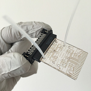
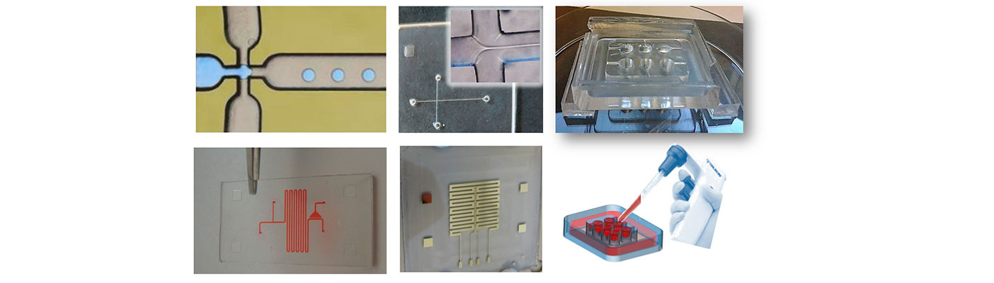
The Lab-on-a-Chip (LoC) and Organ-on-a-chip (OoC) are a wide and multifaced family of miniaturized devices aiming at performing multiple analyses better than conventional methods (LoCs) and recapitulating the essential physio-pathological functions of living human organs (OoCs).
Our current activities in the LoC field are mostly focused on:
- LoC for BioMedicine based on cell chips with an impedance read-out for (i) real-time monitoring of cellular processes such as attachment, micromotion, migration, proliferation and apoptosis. New research directions include application to (ii) sperm cell analysis, the detection of (iii) circulating cells in liquid biopsy approach, and (iv) drug screening.
- LoC for (bio)Chemistry including microfluidic (bio)reactors and Micro Total Analysis Systems (µ-TAS) (i) to produce pharmaceuticals in continuous flow, (ii) to promote cell adhesion/aggregation, (iii) to implement separation/purification of complex fluids, and (iv) to monitor a variety of physical and (bio)chemical processes at microscale.
In the OoC field, our efforts are dedicated to in vitro replication of a miniature patient-on-chip for the development of individually tailored therapies (personalized/precision medicine).
In particular, we develop:
- OoC for neurodegenerative diseases to reproduce (i) the neuro-motor unit and (ii) the blood brain barrier.
- OoC for cancer disease, for a better understanding of tumor dynamics and metastasis (intra- and extravasation) phenomena.
Keywords: In Vitro 3D Culture Systems, Biomaterials Fabrication, Disease Modelling, Drug Screening
People: Anil Chandra, Barbara Cortese, Eliana D’Amone, Stefania D’Amone, Loretta L. del Mercato, Antonio Gaballo, Francesca Gervaso, Giulia Morello, Lorenzo Moroni, Pamela Mozetic, Elisabetta Perrone, Alessandro Polini, Eloisa Sardella, Antonella Stanzione, Alessandra Quarta
We work to produce reliable three-dimensional (3D) cell culture systems as in vitro platforms for cancer and neurological disorders modeling and drug screening assays, with a main current focus on Amyotrophic lateral sclerosis (ALS), pancreatic cancer and glioblastoma.
Our current activities are mostly focused on development of:
- Scaffold-free systems (e.g., spheroids, organoids);
- Thermo-responsive, light-responsive and pH-sensitive polymer-based hydrogels, (e.g., chitosan, pectin, GelMA, alginate and related co-polymers);
- Droplet microfluidics-based high-throughput 3D microgels (e.g., alginate, matrigel, PEG);
- Electrospun polymer fibers (e.g., PCL, PLLA, PLGA);
- Smart biomaterials and 3D constructs with inductive or triggering stimuli to effect cells’ responsiveness to internal or external stimuli (i.e. mechanical and electrical).
The physico-chemical properties of the 3D cell models are characterized by means of standard techniques, such as time lapse CLSM, SEM, XRD and FTIR, DSC and TGA, electrophysiological, mechanical analysis and rheometry. Additionally, the growth and biological features of the 3D models are characterized through morphological and metabolic analysis, expression of surface antigens, and evaluation of cell-cell and cell-microenvironment interactions. We use plasma processed 3D scaffolds to support cell seeding both toward tissue engineering and in vitro platforms for cancer and neurological disorders. Plasma deposition of free-standing nanofilms is studied for applications like cell-sheet and skin engineering.
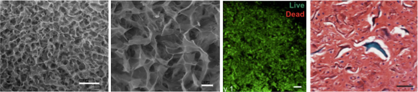
Figure: Collagen scaffold seeded with human mesenchymal stem cells and cultured under chondrogenic conditions (doi.org/10.1016/j.jmbbm.2018.06.040)
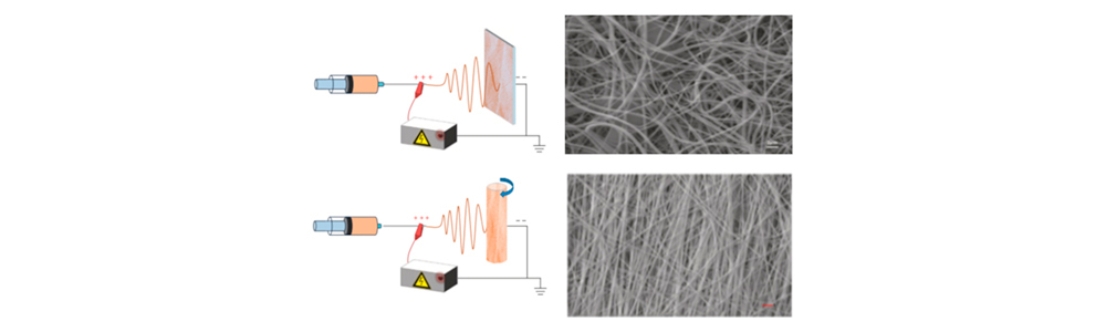
Figure: Chitosan-based mats produced by electrospinning with static (top) and rotating (bottom) collectors for ligament and tendon regeneration (doi.org/10.1155/2018/3651480)
Keywords: Advanced Imaging, Super resolution, Advanced X-ray Tomography, Dynamic Imaging, Multimodal Imaging
People: Silvio Bianchi, Inna Bukreeva, Ivan Colantoni, Barbara Cortese, Loretta L. del Marcato, Michela Fratini, Marco Leonetti, Claudio Maggi, Alessandra Quarta, Filippo Saglimbeni, Ornella Ursini, Ilenia Viola
The Imaging platform is transversal to the research activities and provides skills at the service of specific scientific problems in the nano-biotechnology field.
The principal activities are:
- Advanced fluorescence microscopy. Various fluorescence microscopy techniques such as time-lapse microscopy, confocal microscopy with SIM tornado scanning, FLIM imaging, photoluminescence spectroscopy, multiphoton and STED microscopy are applied to monitor and quantitatively analyze complex dynamic events in living cells and 3D structures (tissues, organoids, scaffolds) at high spatial and temporal rate. Microscopy-based imaging approaches have been developed to observe cellular distributions and movements, in response to cues of the cell microenvironment. Advanced microscopy, in scattering and reflection mode, is also applied for quantitative characterizations of photonic and optoelectronic new materials and devices.
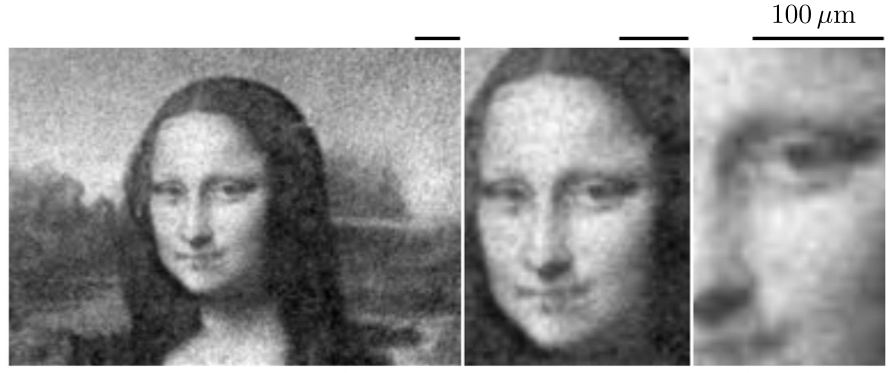
- Super resolution in turbid media. It is a technique facing two problems: on one side allows to study systems in which illumination is deformed by scattering, on the other employs scattering to augment resolution. The “Scattering Assisted Imaging” enables to improve resolution of a factor 2.5.
- X-ray Phase Contrast Tomography. X-ray Phase Contrast Tomography provides resolution and contrast at cellular level also in soft tissues. It makes possible multi-scale 3D biomedical imaging of neuronal and vascular networks ranging from cells through to brain as a whole.
- X-ray Diffraction and Fluorescence. Scanning X-ray micro diffraction (XRmD) exploits a focused sub-micrometer X-ray beam to achieve atomic information with high spatial resolution. It is combined with Scanning X-ray Fluorescence (XRF).
- MRI & Multimodal approach. MRI provides several different methods, including functional imaging, for visualizing tissues. We developed a multimodal approach based on the combination of MRI with high resolution imaging techniques to study the microstructural degeneration in the CNS.
- Holographic microscopy. Digital Holographic Microscopy allows for 3D imaging of colloids/cells from a single 2D snapshot of scattered light interfering with a reference.
- Computer science tools. Development and application of state-of-the-art computer science tools for the effective investigation of 3D object properties.