Joint Seminar - Convolutional neural networks in the segmentation of rodent brain MRI (1), Advanced diffusion MRI: new method to image the complexity of brain (2)
Abstract
(1) Segmentation is a fundamental step in preclinical magnetic resonance imaging (MRI) studies, and these common procedures are still usually performed manually. In this talk, I will outline the potential that deep convolutional neural networks (CNNs) have for these tasks by introducing two case studies (region segmentation in mouse MRI and lesion segmentation in rat MRI).The segmentation models are based on variations of the encoder-decoder network structure. I will present evaluation result with large test-sets of MRIs from hundreds of animals and demonstrate that 1) CNNs outperform multiatlas segmentation strategies in region segmentation, 2) they can be generalized over different phenotypes, and 3) that they can lead to increased reproducibility of lesion segmentation.
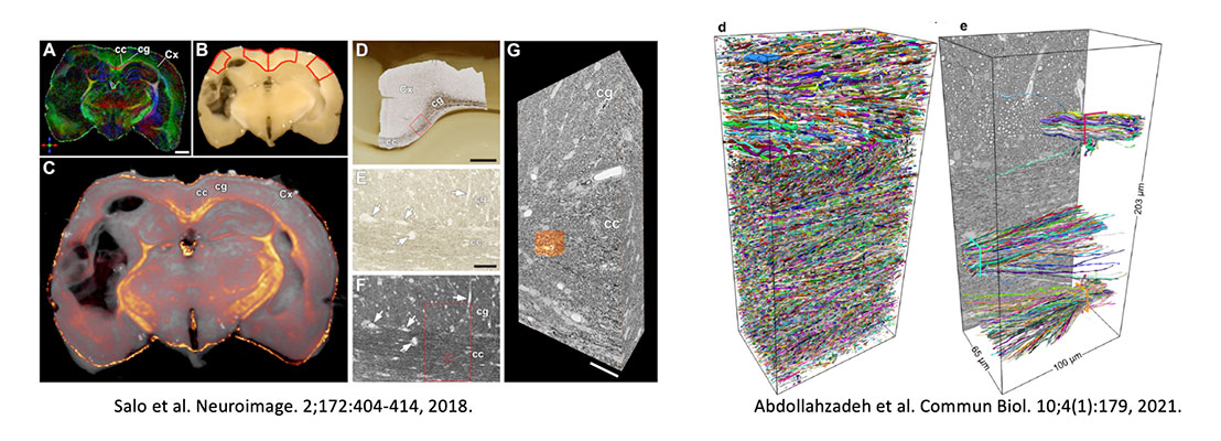
(2) Neuroimaging methodologies, especially magnetic resonance imaging (MRI), have enormously evolved in the recent years. Emerging methods have a great potential to image the brain in a more sensitive and specific manner than conventional ones. However, the challenge is the interpretation of these advanced imaging methodologies in terms of histopathology. In this presentation, I will introduce the advanced diffusion MRI techniques, which improve the non-invasive detection of the healthy and diseased brain. My team works on a multimodal approach including cutting-edge MRI technology, 3-dimensional (3D) histological characterization and computational methods on animal models of brain diseases. The association of MRI and histopathological features is key to understand and interpret non-invasive imaging methods, which can considerably improve the diagnosis, personalized treatment, and/or planning of surgery for patients suffering brain diseases.
Short Bio (2): Alejandra Sierra is a Research Director at A.I. Virtanen Institute for Molecular Sciences (AIVI), University of Eastern Finland (UEF) in Kuopio, Finland. She obtained her Ph.D. in Biochemistry by the Autonomous University of Madrid, Spain (2006). After that she moved to Kuopio in Finland at the Biomedical Imaging Unit (BIU), AIVI, UEF, where she has developed her scientific carrier, first as Postdoctoral Researcher (2010-2012), and also as Research Fellow (2014-2019) from Academy of Finland. Now, she leads the Multiscale Imaging Group focusing on multiscale imaging of the healthy and injured brain. Her special expertise is the combination of magnetic resonance imaging (MRI) techniques with advanced histopathological imaging and analyses methods to understand the tissue contrast produced by different MRI modalities in animal models of brain disease. Research funding from Jane and Aatos Erkko Foundation, Academy of Finland, NIH, Doctoral Program in Molecular Medicine (UEF) alejandra.sierralopez@uef.fi; https://uefconnect.uef.fi/en/group/multiscale-imaging-group/